


08 May 2026 – Modena, Italy
Pterygoid Implants: An Advanced Solution for the Treatment of the Atrophic Maxilla
Pterygoid implants represent an effective fixed implant treatment for the atrophic posterior maxilla. They allow stable anchorage in the pterygoid bone; cortical, compact, not subject to resorption and without the need for bone grafts or sinus lift procedures. In combination with zygomatic, trans-sinus, and trans-nasal implants, they constitute the foundation of the Maxilla-For-All® treatment concept for severe maxillary atrophy.
In the scientific literature, pterygoid implants are widely documented, as demonstrated by several internal JD publications and external studies such as the systematic review by Raouf K, Chrcanovic BR Clinical Outcomes of Pterygoid and Maxillary Tuberosity Implants: A Systematic Review. J. Clin. Med. 2024, 13(15), 4544.
The survival rate is very high but depends on strict adherence to a precise surgical protocol. In the section “Mistakes to avoid” of the book Maxilla-For-All®: Extra alveolar anchorage sites for the rehabilitation of severe maxillary atrophy by Dr. Tommaso Grandi, page 50 of the book discusses the five most critical mistakes with three-dimensional clinical images and their related consequences. This guide analyzes them one by one.
1. Incorrect implant angulation
The trajectory of the pterygoid implant is the most critical variable of the entire procedure. An angulation error compromises cortical engagement and therefore the possibility of obtaining good primary stability.
The book describes three main scenarios:
- Excessive anteroposterior inclination: the implant remains short and anchors only in the tuberosity without reaching the pyramidal process and therefore the pterygoid cortical bone. Insufficient primary stability and impossibility of immediate loading.
- Excessive medial inclination: perforation of the internal pterygoid plate with possible swallowing discomfort for the patient.
- Excessively distal positioning: insufficient posterior bone remains around the implant neck, increasing the risk of tuberosity fracture during implant placement.
It is essential to verify the trajectory already during the first drilling phases, using a radiographic pin and maintaining constant control of the implant direction before proceeding with the following drills.
For further details, you can explore the complete protocol with clinical cases and anatomical references in the book Maxilla-For-All®: Extra alveolar anchorage sites for the rehabilitation of severe maxillary atrophy and the step-by-step clinical videos available on JDentalCare.

2. Inadequate surgical access
Insufficient flap extension is the cause of many subsequent errors. When exposure of the tuberopterygoid region is inadequate, the clinician loses visual and tactile control during drilling, increasing the risk of trajectory deviation, often not immediately visible.
The instrument specifically created for this phase is the JDPterygo Retractor, designed to reach the base of the pyramidal process and guide the correct osteotomy inclination while maintaining flap stability during implant insertion.

3. Incorrect drilling sequence
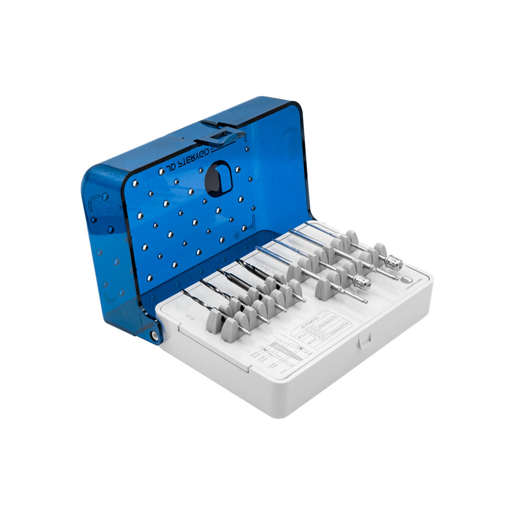
The osteotomy for pterygoid implants, as defined by the JDPterygo Surgical Kit, does not allow variations. Each step prepares the next one: if an intermediate drill is skipped, the following drill works under conditions for which it was not designed, resulting in loss of trajectory control during the most critical phase.
However, there is one challenge that the protocol alone cannot solve: bone quality. In D3/D4 bone, commonly found in the posterior tuberosity, the implant may deviate during insertion, rotating on itself without advancing along the planned trajectory. The temptation may be to increase insertion force. This is a mistake that must be avoided. The correct solution is slow and controlled manual insertion. If the implant does not advance, it should be removed, the trajectory corrected, and the insertion repeated.
4. Lack of three-dimensional CBCT planning
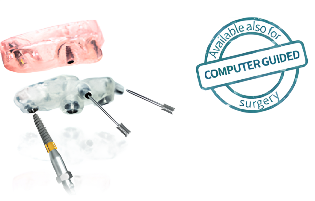
Pterygoid implants can be placed using guided surgery. Performing pterygoid surgery without a preoperative Cone Beam Computed Tomography means operating without knowing the patient’s anatomy. CBCT for three-dimensional evaluation of bone anatomy is the only way to assess two parameters that influence the entire treatment plan and cannot be clinically estimated:
- Bone quality of the tuberosity. Literature highlights a high variability in both bone quantity and quality within the maxillary tuberosity region, with highly heterogeneous dimensional values among patients, making three-dimensional CBCT planning indispensable.
- Position of the pterygopalatine artery. It runs within the pterygopalatine fossa close to the posterior wall of the maxillary sinus: it represents the main vascular risk in pterygoid surgery, especially with implants longer than 30 mm.

What is the solution for safer pterygoid surgery? Guided surgery can help you follow a controlled protocol and reduce deviations.
In the most complex cases there is also the PAGA Concept (Pterygoid Anatomy Guided Approach): computer-guided surgery for the trans-sinus placement of pterygoid implants without trajectory deviations. The implant crosses the maxillary sinus and reaches the pyramidal process following exactly the path planned on the CBCT.
For further reading, JD studies and publications on the accuracy of guided pterygoid implant placement are available together with references from major scientific societies such as the EFP – European Federation of Periodontology.
5. Failure to engage the pterygoid cortical bone
This is the error that compromises all the previous work. You may perfectly plan the case with CBCT, perform flawless surgical access and follow every step of the drilling sequence: if the implant does not engage the pterygoid cortical bone, the technique has failed.
The consequences of failed cortical engagement are immediate:
- Insufficient torque.
- Lack of primary stability.
- Inability to perform immediate loading.
pterygoid implants technique
How to learn and improve your training
Understanding the mistakes is the starting point. Hands-on training is the next step. For both theoretical and practical insights we recommend the book Maxilla-For-All®: Extra alveolar anchorage sites for the rehabilitation of severe maxillary atrophy by Dr. Tommaso Grandi, which includes practical step-by-step videos of real clinical cases.
Additional useful resources:
→ JDentalCare Scientific Publications
Conclusions
Pterygoid implants work; the data proves it. But the outcome depends on five factors: correct angulation, adequate surgical access, an uninterrupted drilling sequence, preoperative CBCT planning, and confirmed engagement of the pterygoid cortical bone. Neglecting any of these variables can transform a highly effective procedure into an early failure.
The pterygoid technique rewards clinicians who train properly and use the right instruments. It penalizes improvisation.
The JDentalCare Team
pterygoid implants technique, pterygoid implants risks
FAQ — Pterygoid Implants
What are pterygoid implants?
Angled implants that reach the pterygoid bone, are cortical, compact and are not subject to resorption; used to rehabilitate the atrophic posterior maxilla without bone grafts. Available in Ø 3.3 mm (JDPterygo One) and Ø 4.0 mm (JDPterygo) versions, used within the Maxilla-For-All® protocol.
What are the risks of pterygoid implants?
The main risks are failure to engage the pterygoid cortical bone, vascular complications involving the pterygopalatine artery in implants longer than 30 mm and tuberosity fracture in case of excessively distal implant positioning.
What is the pterygoid implant technique?
Preoperative CBCT planning, surgical access using the JDPterygo Retractor, progressive drilling sequence with the JDPterygo Surgical Kit and tactile verification of cortical engagement. It can be performed freehand or through guided surgery with PAGA Concept and JD-igital Guide.
What is the PAGA Concept?
Pterygoid Anatomy Guided Approach: a computer-guided surgical technique with a 9 mm sleeve developed by JDentalCare for trans-sinus placement of pterygoid implants without trajectory deviations.
Pterygoid implants technique: an advanced solution for treating the atrophic posterior maxilla without bone grafts or sinus lift procedures.
The pterygoid implant technique is today one of the most effective solutions for rehabilitation of the atrophic posterior maxilla.
Pterygoid implants technique and pterygoid surgery: how to achieve primary stability and immediate loading in cases of maxillary atrophy.
pterygoid implants technique
Pterygoid implant technique: clinical protocol
The pterygoid implant technique requires CBCT planning and trajectory control.